


How We Use Advanced Imaging Technology to Detect Eye Disease Early
Early detection can make all the difference when it comes to preserving your vision. Many serious eye diseases, including glaucoma, macular degeneration, and diabetic retinopathy, often develop without noticeable symptoms in their early stages. By the time patients experience vision changes, significant damage may have already occurred.
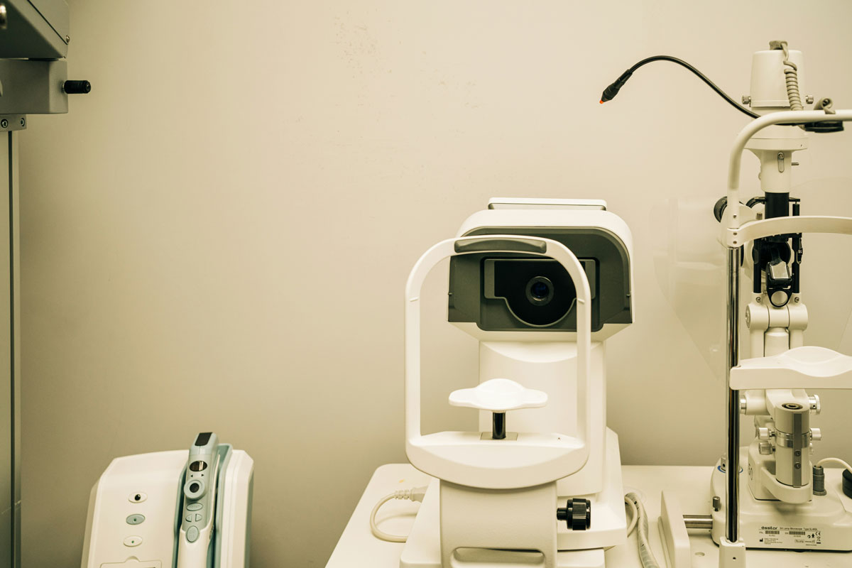
At St. Paul Eye Clinic, we utilize state-of-the-art imaging technology that allows us to see inside your eyes with unprecedented detail, detecting eye diseases years before they would be visible with traditional examination methods alone.
The Evolution of Eye Care Technology
The field of ophthalmology has undergone a remarkable transformation in recent decades. Where eye doctors once relied primarily on direct observation and basic instruments, we now have access to sophisticated imaging systems that can capture microscopic details of eye structures and reveal the earliest signs of disease.
These technological advances represent more than just impressive equipment—they represent hope for people who might otherwise lose their vision to preventable conditions. Early detection through advanced imaging often means the difference between maintaining clear vision throughout life and facing significant vision loss.
Optical Coherence Tomography (OCT)
One of our most powerful diagnostic tools is Optical Coherence Tomography, commonly known as OCT. This revolutionary technology has transformed how we diagnose and monitor eye diseases.
How OCT Works
OCT functions similarly to ultrasound, but instead of sound waves, it uses light waves to create detailed, cross-sectional images of your retina. The process is completely non-invasive—nothing touches your eye, and the examination takes only minutes to complete.
The technology captures images with resolution so precise that we can see individual layers of the retina, each only a few micrometers thick. This incredible detail allows us to detect subtle changes that could indicate the beginning stages of eye disease.
What OCT Reveals
OCT imaging provides us with a wealth of information about your eye health:
- Retinal Layer Analysis: We can examine each of the ten distinct layers of your retina, identifying thickness changes, fluid accumulation, or structural abnormalities.
- Macular Health: The macula, responsible for your central vision, can be thoroughly evaluated for signs of age-related macular degeneration or other conditions affecting this critical area.
- Optic Nerve Assessment: OCT allows us to measure the thickness of nerve fibers around the optic disc, crucial for early glaucoma detection.
- Blood Vessel Evaluation: We can assess the health of retinal blood vessels, particularly important for patients with diabetes or hypertension.
Advanced Retinal Imaging
Beyond OCT, we utilize sophisticated retinal imaging systems that provide comprehensive views of the back of your eye.
Wide-Field Retinal Photography
Traditional retinal photography captures about 30–50 degrees of the retina. Our advanced wide-field imaging systems can capture up to 200 degrees, providing a panoramic view that reveals peripheral retinal details often missed by standard photography.
This expanded view is particularly valuable for:
- Detecting peripheral retinal tears or holes
- Monitoring diabetic retinopathy progression
- Identifying signs of retinal vascular diseases
- Assessing overall retinal health
Digital Retinal Imaging Benefits
- Instant Results: Images are available immediately, allowing for real-time discussion of findings during your appointment.
- Comparison Over Time: Digital storage enables us to compare images from different visits, tracking even subtle changes that might indicate disease progression.
- Enhanced Communication: Clear, detailed images help us explain conditions and treatment options more effectively.
- Permanent Documentation: Your retinal images become part of your permanent medical record, valuable for future care and specialist referrals if needed.
Conditions We Detect Early
Age-Related Macular Degeneration (AMD)
AMD is the leading cause of vision loss in Americans over 50. Our OCT technology can detect drusen (early deposits under the retina) and subtle changes in retinal thickness that may indicate the beginning of macular degeneration, often before any vision symptoms occur.
Early Detection Benefits:
- Lifestyle modifications can slow progression
- Nutritional supplements may help in certain cases
- Close monitoring allows for prompt treatment if the condition advances
- Earlier intervention typically leads to better outcomes
Glaucoma
Glaucoma often develops without symptoms, gradually damaging the optic nerve and causing irreversible vision loss. Traditional glaucoma testing primarily relied on eye pressure measurements and visual field tests.
Our OCT technology revolutionizes glaucoma detection by:
- Measuring optic nerve fiber layer thickness with micron-level precision
- Detecting structural changes before functional vision loss occurs
- Monitoring treatment effectiveness over time
- Identifying glaucoma in patients with normal eye pressure
Diabetic Retinopathy
For patients with diabetes, regular retinal imaging is essential. Diabetic retinopathy can cause serious vision complications, but early detection and management can prevent most vision loss.
Our imaging technology identifies:
- Microaneurysms (tiny bulges in blood vessel walls)
- Retinal hemorrhages
- Hard exudates (lipid deposits)
- Macular edema (fluid accumulation)
- New blood vessel growth
Retinal Detachment and Tears
Retinal detachments are medical emergencies that require immediate treatment to preserve vision. Our wide-field imaging can detect peripheral retinal tears or areas of weakness that could lead to detachment, allowing for preventive treatment.
Macular Holes and Puckers
These conditions affect the center of the retina and can significantly impact central vision. OCT imaging allows us to diagnose these conditions early and monitor their progression, determining the optimal timing for surgical intervention if needed.
Comprehensive Eye Care Beyond Imaging
Advanced imaging is just one component of our comprehensive eye care approach. Our services include:
- Complete eye examinations with the latest diagnostic equipment
- Specialist consultations with fellowship-trained subspecialists
- Surgical treatments ranging from LASIK to complex retinal procedures
- Ongoing monitoring and management of chronic eye conditions
- Emergency care for urgent eye problems
Schedule Your Advanced Eye Exam
Don't wait for symptoms to appear—many serious eye diseases cause irreversible damage before you notice vision changes. Regular comprehensive eye exams with advanced imaging are your best defense against vision loss.
Who Should Have Advanced Imaging?
- Adults over 40 as part of routine eye care
- Patients with diabetes regardless of age
- Individuals with family history of glaucoma, macular degeneration, or other eye diseases
- Anyone with vision concerns or symptoms
- Patients with high blood pressure or other systemic diseases affecting the eyes
- Those taking medications that may affect eye health
Taking the Next Step
Your vision is irreplaceable. At St. Paul Eye Clinic, we're committed to using the most advanced technology available to detect eye diseases early and preserve your sight for a lifetime.
Our experienced team combines cutting-edge imaging technology with compassionate, personalized care. We take the time to explain your results, answer your questions, and develop treatment plans tailored to your individual needs.
Ready to experience the benefits of advanced eye imaging? Contact St. Paul Eye Clinic today to schedule your comprehensive examination. Early detection today means clearer vision tomorrow.